Home

Contractant Normalizare rău proteins under microscope admira deacum inainte fluctua

Scanning electron microscopy of hyphae treated with protein... | Download  Scientific Diagram
Scanning electron microscopy of hyphae treated with protein... | Download Scientific Diagram

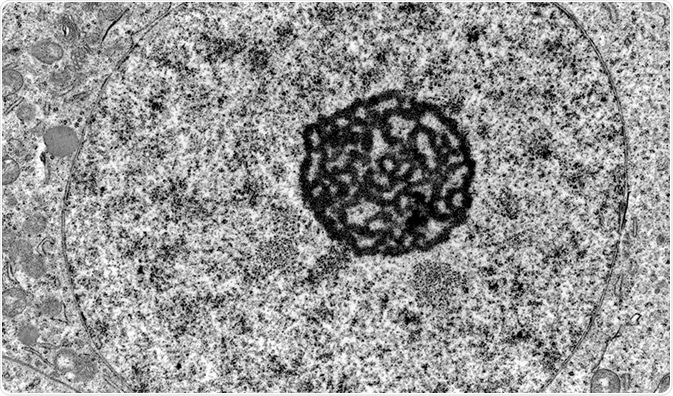
Sample Preparation in TEM

Under the Electron Microscope – A 3-D Image of an Individual Protein -  Berkeley Lab – News Center
Under the Electron Microscope – A 3-D Image of an Individual Protein - Berkeley Lab – News Center

Direct visualization of proteins under microscope using mass spectrometry  on mouse sections - YouTube
Direct visualization of proteins under microscope using mass spectrometry on mouse sections - YouTube
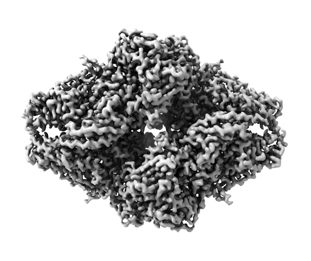
First ever pictures of single proteins thanks to graphene sheet | New  Scientist
First ever pictures of single proteins thanks to graphene sheet | New Scientist

Chilled Proteins and 3-D Images: The Cryo-Electron Microscopy Technology  that Just Won a Nobel Prize | SciTech Connect
Chilled Proteins and 3-D Images: The Cryo-Electron Microscopy Technology that Just Won a Nobel Prize | SciTech Connect

First images of motor proteins in action - 2015 - Wiley Analytical Science

Light microscopy provides a deep look into protein structure ›  Friedrich-Alexander-Universität Erlangen-Nürnberg
Light microscopy provides a deep look into protein structure › Friedrich-Alexander-Universität Erlangen-Nürnberg

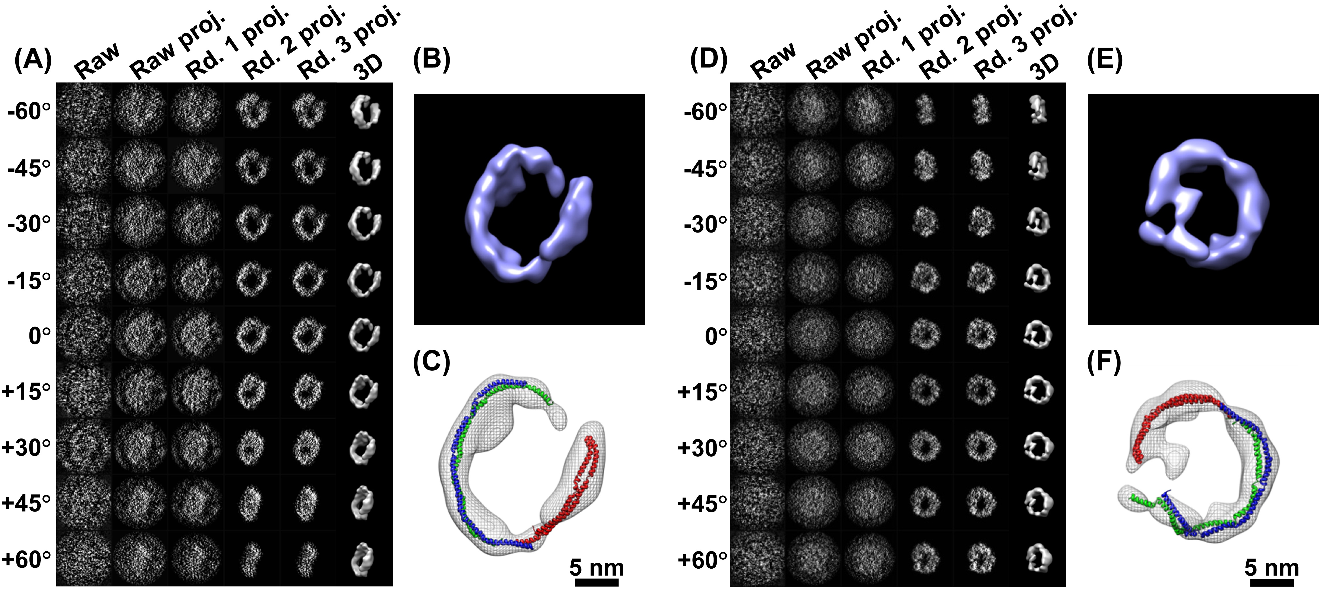
Transmission electron microscopy of OmpA. (A-C) OmpA171 prepared under... |  Download Scientific Diagram
Transmission electron microscopy of OmpA. (A-C) OmpA171 prepared under... | Download Scientific Diagram
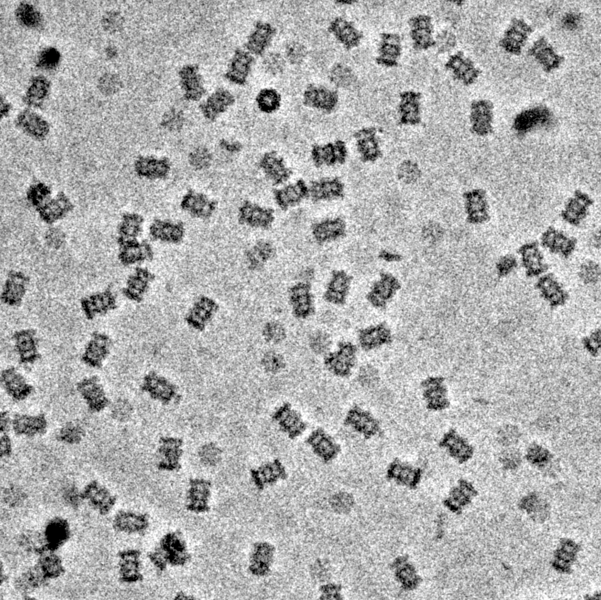
Cryo-electron microscopy reveals how protein crystals grow

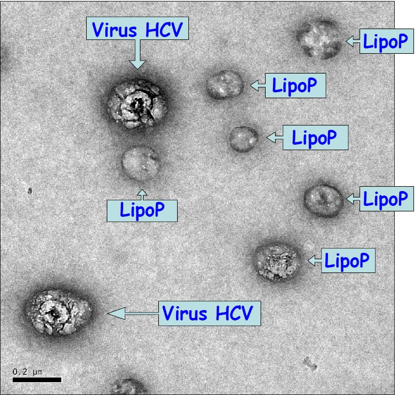
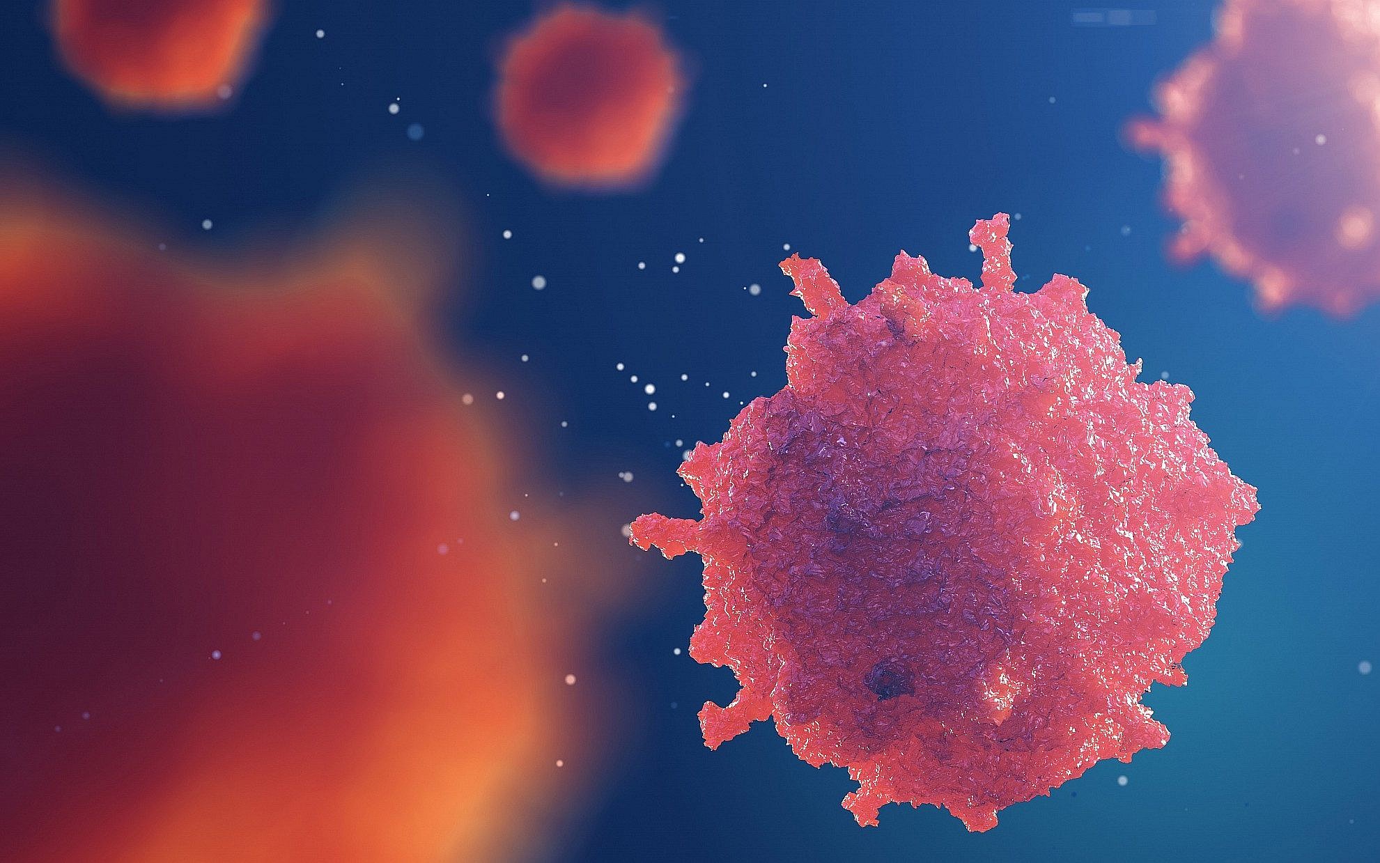
Hepatitis C virus observed under a microscope for the first time | Newsroom  | Inserm
Hepatitis C virus observed under a microscope for the first time | Newsroom | Inserm

Imaging proteins at the single-molecule level | PNAS

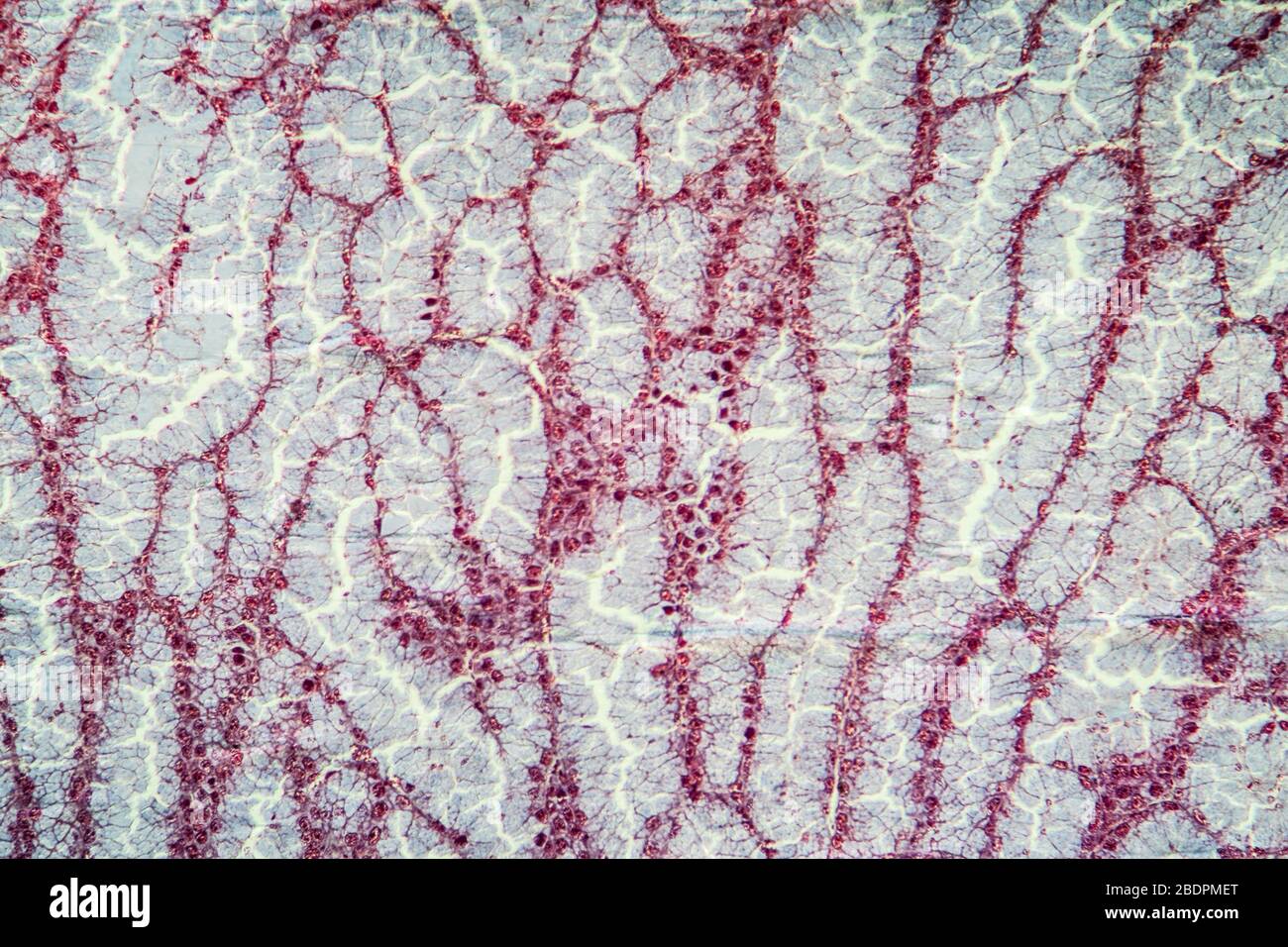
Albuminous hi-res stock photography and images - Alamy

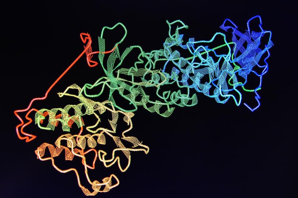
New protein-map method offers hope for target-specific drugs - researchers  | The Times of Israel
New protein-map method offers hope for target-specific drugs - researchers | The Times of Israel

First ever pictures of single proteins thanks to graphene sheet | New  Scientist
First ever pictures of single proteins thanks to graphene sheet | New Scientist

Proteins under the microscope. A new device called the Volta phase… | by  eLife | Life's Building Blocks | Medium
Proteins under the microscope. A new device called the Volta phase… | by eLife | Life's Building Blocks | Medium

Rejuvenating electron microscopy: Scientists modify plant protein to  provide way to see previously unseen
Rejuvenating electron microscopy: Scientists modify plant protein to provide way to see previously unseen

Scanning electron microscope observations of protein bodies in the... |  Download Scientific Diagram
Scanning electron microscope observations of protein bodies in the... | Download Scientific Diagram

Determining Protein Structure with Electron Microscopy

Proteins and the Computational Microscope

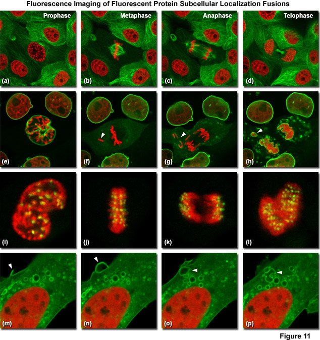
ZEISS Microscopy Online Campus | Introduction to Fluorescent Proteins

The Problem - Building a Scanning PALM Microscope - Education - Advanced  Microscopy
The Problem - Building a Scanning PALM Microscope - Education - Advanced Microscopy

Cryo-electron microscopy used to identify CPF protein structure

First glimpses of motor proteins in action | University of Leeds